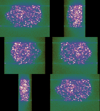
Our group aims to develop optical technologies that allow the imaging of biological systems with high-resolution, correcting for the imaging errors introduced by the sample and subsequently detecting and characterising the processes involved in life.

To do so, we incorporate research on three key technologies:
- Super-resolution imaging allows us to see processes at the nanoscale – much smaller than was generally thought possible only a decade ago. There are multiple approaches to perform super-resolution imaging; we are constructing custom-designed STimulated-Emission Depletion (STED) microscopes for our core research.
- Imaging structures located within biological samples is hampered by the complex nature of the tissue structure. However, it is possible to correct many of the aberrations using adaptive optics. We incorporate dynamic optical elements, such as deformable mirrors and spatial light modulators into our microscopes as a matter of routine.
- By replacing or removing the carbon atoms in a diamond it is possible to make various crystallographic defects. Many of them emit light and some, due to their electronic configuration, are also sensitive to electromagnetic fields. We aim to develop technologies that use the nitrogen-vacancy defect in nanoscopic diamond to detect the activity of neural cells. Due to the requirement to image inside living brains, we will perform the optical measurements in conjunction with the adaptive optic and supperresolution techniques developed in the group.