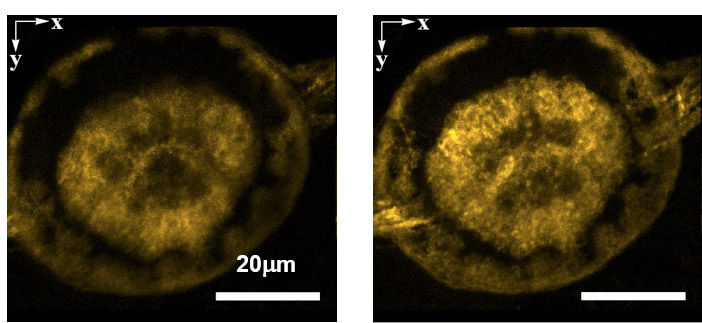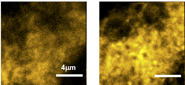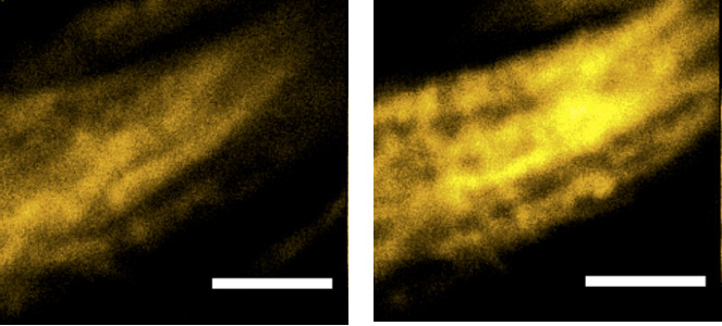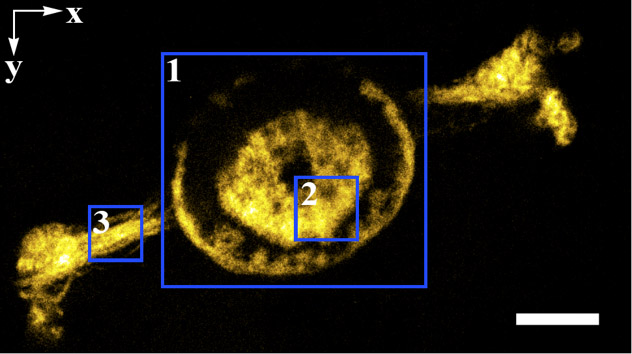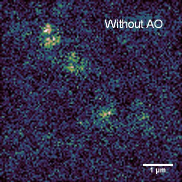
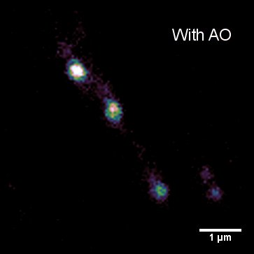
Imaging in biological samples is complicated by the inhomogenous nature of the sample itself – local differences in refractive index distort the light as it propagates and this manifests as aberrations in the resulting image. By incorporating dynamically controllable optical elements, such as deformable mirrors or spatial light modulators, we can correct for many of these aberrations. There are multiple benefits to this approach:
- Improved signal to noise
- We can image at the best resolution for the microscope
- We need less light on the sample – this reduces bleaching and phototoxicity
- We collect more of the light – this is important for samples that will bleach over time or for samples where speed of acquisition is important
Adaptive optics works in a wide range of microscope types – we’re looking at using it in our super-resolution imaging, but it can be applied to most types of microscopes with appropriate choice of hardware!